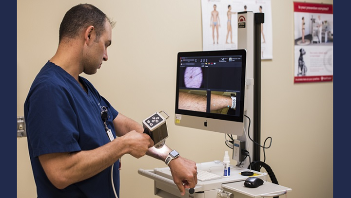
Air Force Maj. Thomas Beachkofsky, 6th Health Care Operations Squadron dermatologist, uses a body scanner microscope to take a picture of a spot on his arm at MacDill Air Force Base, Florida. A new software upgrade allows a complex algorithm to analyze an image captured with a camera and rate the severity of the spot for a dermatologist to review. (U.S. Air Force photo by Senior Airman Adam R. Shanks)
This article is for informational purposes, represents the views of its authors and does not replace professional medical advice. Always consult healthcare professionals for medical advice, diagnosis, or treatment.
Skin cancer is one of the most prevalent forms of cancer in the world, with 16,221 cases being diagnosed annually in Australia alone. The highest risk factor for skin cancer is chronic exposure to UV rays that can cause mutations in DNA and mitochondrial genes. It is further divided into three types; Basal cell, Squamous cell, and melanoma. Of the three, melanoma is the deadliest, as it is responsible for a large proportion of skin cancer-related deaths despite accounting for only 2% of all cancers.
According to the Cancer Council, approximately 2 out of 3 Australians are diagnosed with skin cancer before they turn 70. Recent developments made in the field of skin cancer research have made early diagnosis much easier. Diagnosis of Skin Cancer for any skin pathology involves an initial suspicion for skin cancer due to positive family history and physical examination, after which, definitive diagnosis is made via skin biopsy and histopathological examination.
What is Teledermoscopy?
When a new patient visits a dermatologist for a skin lesion, the dermatologist’s initial step is to examine the lesion with his naked eye. To this end, a device called a dermoscope was designed that facilitates the diagnosis of skin lesions via usage of a magnifying glass and a light source to enhance the visualization of skin lesions in an outpatient setting.
More recently, there has been growing interest in the efficacy of mobile applications that combine the photographic and telecommunication features of a mobile with a magnifying glass to examine skin lesions.
Because it involves the use of telecommunication devices, the device could enable doctors’ to provide consultations to patients living in rural areas eliminating the need for patients living in remote areas to travel long distances for dermatological consultation. But as applications such as these tend to often result in incorrect self-diagnosis by patients, doctors are not quite convinced regarding the benefits of this tool.
Is Self Teledermoscopy equivalent to visiting a dermatologists?
Some researchers who were quite intrigued about the potential clinical applications of teledermatology have recently conducted a study that examines the accuracy of mobile digital teledermoscopy for skin self-examinations.
A randomized, controlled trial done in Brisbane, Australia studied 234 participants who were divided into two groups, the control group, and the interventional group. The participants of the interventional group were provided with iPhone compatible dermatoscopes for conducting self-examinations. Both groups were then asked to perform self-examinations, the controlled group with the naked eye and interventional group with their provided dermatoscopes. Researchers found no significant benefit of either methodology over the other. In fact, the differences in the number of diagnoses between the groups were almost negligible.
The findings of the study revealed no added benefit with the use of teledermoscopy devices for self-examination by patients. The applications were instead found to have both lower sensitivity and specificity in diagnosing skin cancer when compared to naked eye examinations by a dermatologist.
In an online survey performed amongst dermatologists in Australia, researchers found that most medical personnel were open to using dermatoscopy in their practices. They even found it helpful, especially for lesion monitoring. However, they were not very keen on the concept of patients diagnosing themselves with self-assessment tools such as teledermoscopy. The reason for their concern was mainly due to the high risk of a patient discarding malignant skin lesions as being simply benign after self-examination. This can lead to cancer progression and untimely diagnosis of skin cancer when it is at an advanced stage, during which treatment efficacy can be extremely poor.
Although self-examination of skin lesions is still highly recommended by dermatologists, they do not recommend the use of teledermoscopy by patients for diagnosing skin lesions. The scope of self-examination using either a patient’s naked eye or tools such as teledermoscopy should be limited only to the identification of any suspicious skin lesions. Once a suspicious lesion is identified, it should always be followed by a consultation with a dermatologist to rule out skin cancer.
References:
Warshaw E, Greer N, Hillman Y, et al. Teledermatology for Diagnosis and Management of Skin Conditions: A Systematic Review of the Evidence [Internet]. Washington (DC): Department of Veterans Affairs (US); 2010 Jan.
Linares, Miguel A et al. “Skin Cancer.” Primary care vol. 42,4 (2015): 645-59. doi:10.1016/j.pop.2015.07.006
Janda, Monika et al. “Evaluating healthcare practitioners’ views on store-and-forward teledermoscopy services for the diagnosis of skin cancer.” Digital health vol. 5 6 Feb. 2019, doi:10.1177/2055207619828225
Monika Janda, Caitlin Horsham, Dimitrios Vagenas, Lois J Loescher, Nicole Gillespie, Uyen Koh, Clara Curiel-Lewandrowski, Rainer Hofmann-Wellenhof, Allan Halpern, David C Whiteman, Jennifer A Whitty, B Mark Smithers, H Peter Soyer, Accuracy of mobile digital teledermoscopy for skin self examinations in adults at high risk of skin cancer: an open-label, randomised controlled trial, The Lancet Digital Health, Volume 2, Issue 3, 2020, Pages e129-e137, ISSN 2589-7500.
This article is for informational purposes, represents the views of its authors and does not replace professional medical advice. Always consult healthcare professionals for medical advice, diagnosis, or treatment.
Skin cancer is an uncontrolled growth of skin cells resulting from repeated DNA mutations, most often from chronic sun exposure. Despite increasing awareness regarding the risk factors for skin cancer, skin cancer continues to be one of the most prevalent forms of cancers worldwide. Among all the newly diagnosed cases of cancer in Australia, about 80% of the cases are attributed to skin cancer alone. Statistically it has been estimated that approximately one in five individuals in Australia are likely to develop skin cancer during their lifetime.
The two major categories that skin cancers usually fit into include: nonmelanoma and melanoma skin cancer. Nonmelanoma skin cancer involves abnormal growth in keratinocyte – the cell that is found in the outer layer of the skin and produces keratin, while melanoma skin cancer is due to the uncontrolled growth of melanocytes – the cell that produces melanin which gives pigmentation to the skin. Regardless of the type, there are certain risk factors that will increase an individual’s chance of developing either type of skin cancer.
Major Risk Factors For Skin Cancer
Multiple studies have shown irrefutable evidence that ultraviolet radiation from sun exposure, due to its detrimental effect on skin cells, is a major contributing risk factor for skin cancer. Researchers believe that reduction in the thickness of the ozone layer will increase ultraviolet radiation and this will further contribute to an increase in the incidence of melanoma skin cancer. Therefore, overexposure to sunlight can contribute to an increased risk of developing skin cancer.
Additionally, research has indicated that individuals who are fair-skinned are more susceptible to developing skin cancer compared to dark-skinned individuals. Research by Asgari et al. and Berlin et al. has shown that individuals whose family members were diagnosed with skin cancer had an increased likelihood of developing skin cancer in the future.
Furthermore, the number and shape of moles have also been implicated to be a risk factor associated with skin cancer, which has been demonstrated by Heselson in his research confirming the link between the number of moles and melanoma. Fortunately, the measures that individuals need to take that can significantly reduce the risk of skin cancer are quite feasible. With correct guidance and education, the prevalence and incidence of skin cancer can be significantly minimized.
Preventative measures
As previously mentioned, exposure to ultraviolet radiation (UV) from sunlight is the most controllable and important risk factor for skin cancer; therefore, the most critical preventive measure is minimizing exposure to sunlight.
Sun protection such as application of sun protection factor (SPF) of greater than 15 and wearing outfits that cover large body surface areas is crucial, especially for individuals who are fair-skinned or reside in sunny climates.
In addition to protection from sunlight, avoiding indoor tanning is equally important to minimize skin cancer risks. A research conducted by Wehner et al. has shown that out of 9,328 patients with non-melanoma skin cancer, the relative risk for the two most common non-melanoma cancers (squamous cell and basal cell carcinoma) was 1.67 and 1.29 respectively for those who used indoor tanning compared with those who did not perform any indoor tanning. Implementation of programs that educate the general population regarding the prevalence, risk factors, and preventive measures associated with skin cancer is essential to spread awareness about this preventable yet fatal disease.
Prognosis and Survival
The prognosis of skin cancer is complex, multifactorial, and largely dependent on the type, location, and stage of cancer. Non-melanoma skin cancer can have a 5-year survival rate between 95-99%. According to the Skin Cancer Foundation, approximately 5,000 people die monthly from non-melanoma skin cancers.
National Cancer Institute has stated the 5-year survival for melanoma skin cancer between 2014-2016 to be around 92.7%. Overall around the world, the mortality rates of skin cancer are declining. The decline in the mortality rates of skin cancer is mainly due to an upsurge in the number of early diagnoses allowing timely management and prevention of cancer metastasis.
Do all skin lesions need to be biopsied?
If you notice a newly formed skin abnormality that was previously non-existential such as moles, lesions, or spots, it is always best to have an expert examine you. However, in rare instances even though you may not notice any abnormal lesion, it might be worthwhile to visit a dermatologist to assess your risks if one of your family members has been diagnosed with skin cancer. According to the National Cancer Institute, simple screening tests by a general practitioner for skin cancer have not yet shown to reduce your likelihood of dying from skin cancer. Trials are currently underway to examine the effect of screening tests on survival outcomes and mortality. Whenever in doubt regarding any abnormality in your skin, it is highly recommended to visit a dermatologist, where you can be examined thoroughly and in some cases may be advised to undergo a skin biopsy if the dermatologist suspects skin cancer.
Skin biopsy: The Gold Standard
Currently, a skin biopsy is a gold standard for the diagnosis of skin cancer. Skin biopsies are simple procedures usually performed in an outpatient setting and take less than 20 minutes.
What does a skin biopsy involve?
The first step in skin biopsy involves sterilization of the site with an alcohol swab and application of a local numbing agent. There are various types of skin biopsies including incisional, excisional, punch, and shave; the dermatologist will opt for an appropriate type of skin biopsy based on a multitude of factors associated with the lesion.
After the biopsy is completed, depending on the type of incision, a stitch may be needed at the site of the biopsy. Patients need to be careful to keep the area sterile and moist in order to prevent infection. Typically, a small scar may be visible after a biopsy.
How is the biopsied tissue examined?
The tissue obtained from a biopsy is sent to a laboratory where it is examined and tested to identify any pathologic evidence of disease. The time it takes for the results to come back vary by institution, however, it usually takes approximately one to two weeks for the pathologist to report the results and send them to your dermatologist. Once the results are sent to the dermatologist, a final diagnosis will be made.
What will be the next step in case the skin biopsy is positive for cancer?
If the skin biopsy report confirms skin cancer, the dermatologist will discuss treatment plans with the patient. Occasionally, it may be necessary to conduct other tests to determine the stage of skin cancer (Stage I to IV i.e., early to advanced stages). Treatment options include immunotherapy, targeted therapy, cryotherapy, chemotherapy, and surgical removal of cancerous tissue. Mohs surgery may be an option in early skin cancer, in which layers of cancerous cells are removed and examined under the microscope until no cancerous tissues remain.
Early detection For Better Survival
Early detection is a pivotal strategy to increase survival rates and lower mortality in all types of cancer. Therefore, performing skin biopsies early on can increase the chances of identifying the disease. A study by Welch and colleagues published in BMJ showed that an increase in the biopsy rates was associated with an increase in the incidence of melanoma skin cancer between 1986 and 2001. This illustrates the importance of skin biopsies as not merely a tool for diagnosis of skin cancer, but also for reducing mortality rates by early diagnosis and appropriate therapy.
Likewise, a study by Skaggs and Coldiron demonstrated an increase in the use of skin biopsies by 153% between 1993-2016 while treatment for skin cancer has only increased by 39% within the same timeframe. Thus, this shows potential in skin biopsies as being both a diagnostic and treatment option for skin cancer.
A delay in the detection of skin cancer has been shown to significantly affect the 5-year survival rate. Conic et al. showed that for patients who had stage I melanoma but waited more than 1 month after biopsy had a 5% worse survival rate compared to 41% worse survival rate in those who waited more than 4 months.
Thus, it is imperative to counsel patients to screen themselves and identify any suspicious lesions. As such there have been many campaigns, one of which is the SPOT Skin Cancer campaign which has directed its efforts at improving patient awareness. Patients can employ the ABCDEF checklist which combines the ABCDE (asymmetry, irregular border, color, diameter, and evolving) and the “ugly duckling sign” (a spot that is different to others) in identifying suspicious lesions.
Skin cancer continues to rise exponentially and has become one of the most prevalent cancers in the world. Therefore, it is valuable to have knowledge concerning risk factors and preventive measures for skin cancer. Moreover, early diagnosis and treatment are vital to increase survival rates and decrease mortality. Skin biopsy has become the most used diagnostic tool for cancer in the past decade as it is non-invasive, relatively safe, and produces quick results. Additionally, it is also being used for the treatment of non-melanoma cells where the cancerous tissue can be removed via core excision. People need to continue to be vigilant in self-screening using the ABCDEF rule as a guide for early detection. Many clinical trials are currently underway to investigate many factors related to skin cancer and possible novel treatment options.
References
Urbach, F. (1980). Ultraviolet radiation and skin cancer in man. Preventive Medicine, 9(2), 227–230. doi:10.1016/0091-7435(80)90080-8
HESELSON J. Moles and melanomas of the skin. S Afr Med J. 1961;35:1113-1120.
Berlin NL, Cartmel B, Leffell DJ, Bale AE, Mayne ST, Ferrucci LM. Family history of skin cancer is associated with early-onset basal cell carcinoma independent of MC1R genotype. Cancer Epidemiol. 2015;39(6):1078-1083. doi:10.1016/j.canep.2015.09.005
Asgari MM, Warton EM, Whittemore AS. Family history of skin cancer is associated with increased risk of cutaneous squamous cell carcinoma. Dermatol Surg. 2015;41(4):481-486. doi:10.1097/DSS.0000000000000292
Brash DE. Sunlight and the onset of skin cancer. Trends Genet. 1997;13(10):410-414. doi:10.1016/s0168-9525(97)01246-8
Wehner Mackenzie R, Shive Melissa L, Chren Mary-Margaret, Han Jiali, Qureshi Abrar A, Linos Eleni et al. Indoor tanning and non-melanoma skin cancer: systematic review and meta-analysis BMJ 2012; 345 :e5909
Skaggs R, Coldiron B, Skin Biopsy and Skin Cancer Treatment Utilization in the Medicare Population, 1993-2016, Journal of the American Academy of Dermatology (2020), doi:https://doi.org/10.1016/j.jaad.2020.06.030.