Categories
research Uncategorized

Why the ABCDE rule is not helpful but dangerous in skin cancer prevention


This article is for informational purposes, represents the views of its authors and does not replace professional medical advice.

The most dangerous and aggressive skin cancer, malignant melanoma, can have a wide variety of appearances and usually develops rapidly. The ABCDE rule (also known as the ABCD rule in some countries) has therefore been promoted to help lay people recognize when it is time to see a dermatologist. But how effective is this rule? Our data clearly shows that another self-monitoring protocol has a superior sensitivity and better outcomes than the outdated ABCDE rule.

Authors: Dr. Tudor, Adrian;  Dr. Feldman, Jonathan; Dr. Diamandis, Carolina – Full research report available here: https://zenodo.org/record/5731554

Status Today

The standard care in the G20 countries has been running for years according to the same scheme: the patient is screened by a dermatologist at certain intervals, usually once a year, and the rest of the time the ABCDE rule2 (regionally slightly different in the interpretation)8 should be used by the patients to detect skin cancer by means of self-observation.

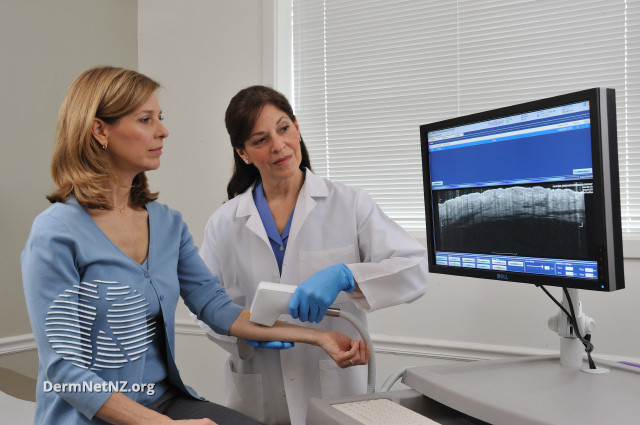
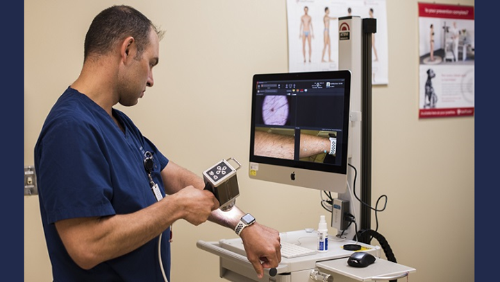
Dermatologists tend to use the dermatoscope rather than the scalpel, just as if an excisional biopsy were a major surgical event, which of course it is not. Instead endless follow-up checks and an absurdly complex system of self-monitoring is imposed on complete laymen:2,3,4,9

The ABCDE rule

A for asymmetry

New skin protocol

Uneven, asymmetrical shape. This means a structure that is not uniformly round, oval or elongated.


B for border:

The margins appear washed out, jagged or uneven and rough. Or a colored skin mark has tongue-like extensions and indentations, blurred contours or grows frayed into the healthy skin area.

C for color

The skin mark is irregular and inconsistently colored, sometimes ranging from jet black to skin colored. Lighter and darker patches can be seen in a mark. The color of the skin mark mixes with pink, gray, blue, or black dots, or it has a crusty overlay.

D for diameter:

Uneven, asymmetrical shape. This means a structure that is not uniformly round, oval or elongated.

The margins appear washed out, jagged or uneven and rough. Or a colored skin mark has tongue-like extensions and indentations, blurred contours or grows frayed into the healthy skin area.

The skin mark is irregular and inconsistently colored, sometimes ranging from jet black to skin colored. Lighter and darker patches can be seen in a mark. The color of the skin mark mixes with pink, gray, blue, or black dots, or it has a crusty overlay.

E for elevation or evolution

If the lesion measures more than five millimeters at its widest point, this is considered critical. However, the exact size specifications differ in the scientific literature. Some propose to replace “diameter” with “dark color”8 which is hard to understand why when color is already included and people with light skin are the minority on this planet. In about 80% of the world population normal nevi are black. Therefore the proposal by Goldsmith and Solomon8 would, if at all, only help caucasian patients. Some might considers this racist, inappropriate and not helpful.

_______

Many medical professionals believe that this complex list can be used by laymen while many physician themselves struggle with it.5 However, the main problem is: countless types of benign skin lesions also exhibit some of these features, in fact almost all lesions have at least one of the “ABCDE” aspects and are thus considered to require diagnostic workup (dermatoscopy or biopsy). On the other hand, many melanomas appear visually like a harmless nevus, bruise, or sore, especially if they develop from an existing nevus or are localized in a difficult-to-see location. This raises the question of how helpful the ABCDE rule is when even many general practitioners have no clue how to use it.5,7

Starting point for us was the subjective impression that two aspects are highly problematic in dermatological daily routine in practices and clinics and yet continue to be applied worldwide:

1)

Regular screening without education about the highly aggressive and fast-growing nature of particularly dangerous skin cancers creates a false sense of security among patients (“illusion of interval safeness”). Vigilance appears to drop dramatically due to this illusory sense of security, as the normal patient associates screening with a reduction in their risk of developing skin cancer, especially melanoma. This is an implicit misleading of all medical laymen, as melanomas can arise de novo at any time and develop rapidly. If studies and practical experience are taken together, patients should actually be informed that a precautionary interval screening is well suited to the early detection of certain types of skin carcinomas, but that this is by no means the case for the classic and rightly feared malignant melanoma, which develops at a very rapid pace. In relation to this most dangerous skin cancer, the annual or otherwise timed interval screening by a dermatologist is mostly useless.3,7,9

2)

The ABCD(E)8 rule in all its regional variants flushes the wrong people into practices and clinics. The rule seems far too complicated and misleading for laymen.3 People over the age of 50, especially those with advanced seborrheic keratoses, fill the waiting rooms because this invariably benign (harmless) entity usually violates almost every letter of the ABCD(E) rule. In contrast, all those whose small, round, sharply marked melanoma “only” bleeds briefly in between stay at home. “Can happen”, the layman thinks, because in fact millions of people every day injure some skin lesion when removing body hair.

Therefore, we were grateful that a befriended research team from Spain provided us with anonymized data from a study that could not be completed there due to a stop in funding.3,5

The details of the study (data and results) can be referred to in the full paper: (No paywall): https://zenodo.org/record/5731554

Discussion

In research, it is rare that study results are so unambiguous. We immediately drew consequences for the management of our patients and introduced a new diagnostic protocol for the prevention of advanced skin cancers of all types, including malignant melanoma. This means: of course we continue to provide our patients with the usual annual interval examinations. However, we have completely abolished the ABCD(E)

rule and replaced it with the C-Rapid-H-Plus Protocol to be used by the patients at home.

C stands for Change in any way, shape, or form.

Rapid means immediate excision without dermatoscopy prior to excision.

H is about leaving to make the definitive diagnosis to the histopathologists.

Plus indicates the offer to send in photos of skin lesions anytime (24/7) during the interval along with a message to be seen, analyzed and responded to by a dermatologist no later than 24 hours.

Since we did launch the C-Rapid-H-Plus Protocol in 2020 in the clinics we work at, it is yet too early to publish reliable data from clinical practice. However, the number of skin cancer cases requiring follow-up treatment after a wide and deep excision has dropped to zero in our own patients. This is remarkable. We assume that it is also related to the less complex patient education and the digital consultation room that is always open. The latter in particular seems to be of immense importance.

Of course, this study has countless limitations. We received the Spanish data only as an anonymized data donation. Even though we know that it was done correctly, we lacked the possibility to ask for some details. Nevertheless, we decided to publish this work because its topic is of profound importance for general health and thousands of relevant situations that occur every day in clinics and practices around the world.

Conclusion

By replacing the ABCDE rule with the C-Rapid-H-Plus Protocol, we were able to reduce the number of advanced skin cancer cases in our patients to zero. A result that is consistent with the data from Spain. Telemedicine will and must also play an important role in this new approach, just as the hesitation against the routine use of scalpels must come to an end.

Conflicts of Interest Dr. Diamandis works in the field of dermatology as a clinician. Dr. Tudor and Dr. Feldman have nothing to declare.

References

  1. Suppa M, Daxhelet M, del Marmol V. Dépistage du mélanome [Melanoma secondary prevention]. Rev Med Brux. 2015 Sep;36(4):255-9. French. PMID: 26591309.
  2. Maire C, Vercambre-Darras S, Desmedt E. Diagnostic du mélanome [Diagnosis of melanoma]. Rev Prat. 2014 Jan;64(1):61-8. French. Erratum in: Rev Prat. 2014 Mar;64(3):349. PMID: 24649548.
  3. De Giorgi V, Papi F, Giorgi L, Savarese I, Verdelli A, Scarfì F, Gandini S. Skin self- examination and the ABCDE rule in the early diagnosis of melanoma: is the game over? Br J Dermatol. 2013 Jun;168(6):1370-1. doi: 10.1111/bjd.12250. PMID: 23738643.
  4. Garrido AQ, Wainstein AJA, Brandão MPA, de Vasconcellos Santos FA, Bittencourt FV, Ledsham C, Drummond-Lage AP. Diagnosis of Cutaneous Melanoma: the Gap Between the Knowledge of General Practitioners and Dermatologists in a Brazilian Population. J Cancer Educ. 2020 Aug;35(4):819-825. doi: 10.1007/ s13187-020-01735-z. Erratum in: J Cancer Educ. 2020 May 27;: PMID: 32193871.
  5. Benelli C, Roscetti E, Dal Pozzo V. Reproducibility of the clinical criteria (ABCDE rule) and dermatoscopic features (7FFM) for the diagnosis of malignant melanoma. Eur J Dermatol. 2001 May-Jun;11(3):234-9. PMID: 11358731.
  6. Coups EJ, Manne SL, Stapleton JL, Tatum KL, Goydos JS. Skin self-examination behaviors among individuals diagnosed with melanoma. Melanoma Res. 2016 Feb;26(1):71-6. PMID: 26426762.
  7. Goldsmith SM, Solomon AR. A series of melanomas smaller than 4 mm and implications for the ABCDE rule. J Eur Acad Dermatol Venereol. 2007 Aug;21(7):929-34. doi: 10.1111/j.1468-3083.2006.02115.x. PMID: 17659002.
  8. Bandic J, Kovacevic S, Karabeg R, Lazarov A, Opric D. Teledermoscopy for Skin Cancer Prevention: a Comparative Study of Clinical and Teledermoscopic Diagnosis. Acta Inform Med. 2020 Mar;28(1):37-41. doi: 10.5455/ aim.2020.28.37-41. PMID: 32210513; PMCID: PMC7085326.

Tudor, Adrian, Feldman, Jonathan, & Diamandis, Carolina. (2021). Why the ABCDE rule is not helpful but dangerous in skin cancer prevention. Zenodo Publishing. https://doi.org/10.5281/zenodo.5731554

Categories
Uncategorized

Differentiating Benign Skin Lesions from Skin Cancer


This article is for informational purposes only and does not replace professional medical advice.

A new skin lesion that you had never noticed before is always a reason for concern. This concern can in select cases be lifesaving owing to the timely diagnosis and treatment of otherwise fatal malignancies. Everyone needs to be constantly vigilant and aware of any suspicious appearing skin lesions due to the poor prognosis associated with a delayed diagnosis of skin cancer. But what is the description of a suspicious skin lesion? Does every mole, freckle and blemish need a skin biopsy to rule out the possibility of a malignant tumor? No, it doesn’t. More often than not, a freckle is just a freckle and will require no further management.

Therefore, any new skin lesion needs to be examined by a professional before jumping to conclusions with a self-assumed diagnosis. To this end the question again arises, what types of skin lesion need a professional opinion? In order to understand this, you will need to know the typical warning signs of a premalignant or malignant skin lesions, which will be discussed below.

Typical Morphological Characteristics of a Malignant Skin Lesion

Size: A new or previously existing skin lesion that changes in size either gradually or rapidly over a period of weeks to even years can be a sign of malignancy. If a new skin lesion is greater than 5mm in diameter, it is best to have it examined.

Appearance: The lesion appears irregular and doesn’t conform to typical feature of moles or freckles. Abnormal non-uniform color, structure and surface of the skin lesion are typical features of a malignant lesion. Multiple skin lesions of new onset with abnormal morphology is an important warning sign that needs to promptly be examined.

Keratin Plug: Keratin plugs can be a benign finding but presence of a large keratin plug over nodules greater than 5 mm in diameter with a suspicious appearance demands a visit to a dermatologist.

Ulcerations or Crust: Skin lesions with spontaneous development of crusts or ulcerations without history of any trauma or irritation is another red flag that needs urgent evaluation.

Adherent Scale: Scales are not a finding of a normal healthy skin, and can be caused by a number of genetic, allergic or autoimmune etiologies. Sometimes, it can also be a sign of a premalignant skin lesion such as actinic keratosis, Bowen disease, lichen planus, etc.

Erythematous Halo: A ring or halo of redness around a nodule or papule resembling a bulls eye pattern can also be seen in malignant skin lesions and therefore needs to be further examined.

Location of the lesion: Although skin cancer can appear in any part of the body, some areas are more prone to develop malignant lesions such as the scalp, peri-ocular region, genital orifices, ungual area, and orifices. Lesions in these regions always require a specialist’s eye for evaluation.

Growth Pattern: Rapidly growing aggressive growth pattern warrants urgent evaluation.

Distinguishing Features Of Skin Cancer Subtypes

Skin Cancer is a broader term for various cancerous skin lesions including many subtypes. Each of them exhibit their own unique distinguishing features such as:

Melanoma

Melanoma is a cancer of the melanocytes present in the basal layer of the epidermis. Its features can be remembered with the easy mnemonic ABCDE

A– Asymmetry
B– Border Irregularity
C– Color Variation
D– Diameter greater than 6mm
E– Evolving

Often, early lesions of melanoma are ignored by the patient due to their similarity in appearance with freckles and moles. All are pigmented and appear benign and can only be differentiated via a biopsy in the early stages. Any mole that display the ABCDE characteristics needs to be evaluated. It should be noted that previously benign mole or freckle may undergo malignant transformation as well.

Squamous cell carcinoma

Squamous cell carcinoma is a cancer of the epidermis which is made of stratified squamous epithelium. They often develop in the form of a plaque or a nodule over a period of weeks to months and can become ulcerated. Painful ulcerated nodules should raise the suspicion of squamous cell carcinoma.

Basal Cell Carcinoma

Basal cell carcinoma involves the basal layer of the epidermis, and usually presents with a typical nodulo-ulcerated lesion with early bleeding. Its growth is slower than squamous cell carcinoma and it can take years for the patient to seek an expert’s opinion due to their relatively slower growth pattern. Therefore, any persisting skin lesion that doesn’t go away within a few weeks should be examined regardless of the growth rate.

Morphological Features of a Benign Skin Lesion

To further aid you in differentiating benign from malignant skin lesions, it is imperative to also know the features of a benign skin lesion.

Size: The size remains constant, stable and grows either very slowly or not at all over decades.

Appearance: Uniform color, shape and structure with no ulcerations, plaques, bleeding or Erythematous ring.

Stability: The size, shape, color and structure of a benign skin lesion doesn’t change over time.

The typical features described above are as stated: typical i.e. they are commonly seen features of malignancy, but atypical cases may not present in a similar manner. Furthermore, all the above features are not a criterion for diagnosis as some malignant skin lesions may present with only one of the above features or in atypical cases none of the above features.

In high risk people such as people with frequent sun exposure, elderly people, smokers, or with genetic predisposition to malignancy, all skin lesions should be considered to have malignant potential until examined thoroughly by an experienced dermatologist familiar with skin cancer.


References

Cancer 2014 Sep 03;[EPub Ahead of Print], VR Belum, AC Rosen, N Jaimes, G Dranitsaris, MP Pulitzer, KJ Busam, AA Marghoob, RD Carvajal, PB Chapman, ME Lacouture

Ribas A, Flaherty KT. BRAF targeted therapy changes the treatment paradigm in melanoma. Nat Rev Clin Oncol. 2011; 8: 426‐ 433.

Jaimes N, Zalaudek I, Braun RP, Tan BH, Busam KJ, Marghoob AA. Pearls of keratinizing tumors. Arch Dermatol. 2012; 148: 976.

Rosendahl C, Cameron A, Argenziano G, Zalaudek I, Tschandl P, Kittler H. Dermoscopy of squamous cell carcinoma and keratoacanthoma. Arch Dermatol. 2012; 148: 1386‐ 1392.

Lacouture M, Chapman PB, Ribas A, et al. Presence of frequent underlying RAS mutations in cutaneous squamous cell carcinomas and keratoacanthomas (cuSCC/KA) that develop in patients during vemurafenib therapy [abstract]. J Clin Oncol. 2011; 29(suppl): 8520.


Categories
Uncategorized

Diagnostic Accuracy of Optical Coherence Tomography in Skin Cancer


This article is for informational purposes only and does not replace professional medical advice.


Over the past decade, Optical coherence tomography (OCT) has been increasingly used as a novel noninvasive imaging device to diagnose skin cancer. Its popularity is attributed to its ability to examine the skin in real time by providing a cross-sectional imaging of the skin. In comparison to older tools used by dermatologists such as the reflectance confocal microscopy, OCT has significantly better imaging depth of up to 2mm and a wider field of view albeit with a lower cellular resolution.

Various forms of OCT have since emerged such as Frequency domain OCT, dynamic OCT (D-OCT), and high-definition OCT (HD-OCT), all of which have been found to be efficacious in the diagnosis, treatment planning as well as monitoring of non-melanoma skin cancers.

With any diagnostic tool, continuous development and assessment is essential to examine the potential of the tool in clinical scenarios. To examine the diagnostic potential and accuracy of a tool, the most important factors are its sensitivity and specificity. However, the accuracy of the OCT is a subject of debate among dermatologists as many have expressed concern that screening with OCT alone as a diagnostic tool may lead to false negatives and delayed diagnosis of skin cancer. With this concern in mind, a research was conducted that examined the diagnostic accuracy of OCT to diagnose skin cancer.

The chief objective of this study was to estimate the diagnostic accuracy of OCT in basal cell carcinomas (BCC) and actinic keratosis (AK) as well as differentiating these lesions from normal skin.

Study Methodology

The study prepared a set of 142 OCT images that met the predetermined selection criteria for image quality as the diagnosis accuracy was heavily dependent on image quality. The OCT images for the study were all obtained by a widely used and commercially available OCT machine. The OCT images contained a mixture of cases with diagnosis of AK, BCC and normal skin.

These images were then presented simultaneously to two groups of blinded observers. One group comprised of 5 dermatologists who were experienced in OCT-image interpretation and diagnosis with a minimum of one-year experience in using OCT. The second group consisted of 5 dermatologists who were unfamiliar with OCT as a diagnostic tool. Each group of dermatologists were asked to answer a standardized questionnaire based on the OCT images and their presumed diagnosis.

Results of The Questionnaire

In the first group of dermatologists who were experienced with OCT, the diagnostic accuracy was found to be at a satisfactory level for Basal Cell Carcinoma with a sensitivity of 86% to 95% and a specificity of 81% to 98%. Unsurprisingly, the second group of dermatologists were not as adept at OCT as a diagnostic tool for BCC with the experienced dermatologists exhibiting an overall higher diagnostic accuracy in comparison to the OCT inexperienced dermatologists.

Proper OCT training is essential early on during residency years for dermatologists to be well versed with OCT, and only then can OCT be an effective screening asset for skin cancer.

References

Olsen J, Themstrup L, De Carvalho N, Mogensen M, Pellacani G, Jemec GB. Diagnostic accuracy of optical coherence tomography in actinic keratosis and basal cell carcinoma. Photodiagnosis Photodyn Ther. 2016 Dec;16:44-49. doi: 10.1016/j.pdpdt.2016.08.004. Epub 2016 Aug 9. PMID: 27519350. Feature image from DermNet, https://creativecommons.org/licenses/by-nc-nd/3.0/nz/legalcode,

Categories
Uncategorized

A 45-Year Historical Cohort Study of Cutaneous melanoma in 14·9 million people


This article is for informational purposes only and does not replace professional medical advice.

In the Nordic countries, the incidence of melanoma has been rising at a gradual yet steady rate in the past century. Multiple smaller scale studies have previously reported an increase in age-adjusted incidence of cutaneous melanoma (CM) in the Nordic countries over the last 60 years. However, there have been few prospective population-based studies that examined the occupational variation in CM risk over a period of time. In order to understand and determine the variations in the incidence of Cutaneous melanoma, a large-scale cohort study was done beginning from 1961 to 2005 by the Nordic Occupational Cancer Study team.

The chief objective of this cohort study was to determine the occupational variation in Cutaneous Melanoma and the associated risk with various occupations.

Study Methodology

Researchers undertook a massive 45-year follow-up of a historical prospective cohort study beginning from 1961 to 2005. The information for the study was obtained by linking records between the population census and cancer registry data for Nordic residents between the ages of 30 and 64 years living in Denmark, Finland, Iceland, Norway and Sweden. The study stratified the occupations into 53 categories each of which was further designated into indoor, outdoor and mixed work to estimate sun exposure. Furthermore, the data also analyzed the socioeconomic status of the entire population to determine its role on cutaneous melanoma.

With this data, standardized incidence ratios (SIRs) were calculated for each category of occupation and socioeconomic status. The SIR was calculated by dividing the observed number of Cutaneous Melanoma cases by the expected number of cases. Data for this calculation was obtained using stratum-specific person-years and national Cutaneous Melanoma incidence rates. The higher the Standardized incidence ratio, the higher should be the risk for skin cancer in a particular occupation.

Study Findings

The study evaluated a total of 385 million person-years, among which 83 898 incident cases of Cutaneous Melanoma was found. The study found that men with outdoor work had a low SIR of 0·79 and men with indoor work had a high SIR of 1·09. The differences in women based on their occupation also found similar results as in the men with higher SIR. In the group categorized under high socioeconomic status, the risk for melanoma was found to be even higher based on their SIRs which was found to be 1.34. in men and 1.31 in women. Likewise, people who worked in military, transport, technical, and public safety workers with substantial skin exposure to carcinogens had higher risk for skin cancer.

Conclusion

The major flaw in the study was the use of SIRs to estimate skin cancer risk as the denominator in the SIR equation was based on an expected number of cases which can be substantially higher when wrongly estimated skewing the equation to the wrong end. In people with outdoor occupation, there was reportedly a higher number of cases of melanoma in comparison to indoor worker but due to the observed cases being higher than the expected cases their SIRs were calculated as low. However, the study did find that occupational and socioeconomic variation in melanoma played a significant role in terms of risk for developing this fatal condition.

References:

Alfonso JH, Martinsen JI, Weiderpass E, Pukkala E, Kjærheim K, Tryggvadottir L, Lynge E. Occupation and cutaneous melanoma: a 45-year historical cohort study of 14·9 million people in five Nordic countries. Br J Dermatol. 2020 Jul 6. doi: 10.1111/bjd.19379. Epub ahead of print. PMID: 33026672.

Categories
Uncategorized

New and Ongoing Research in The Field of Skin Cancer


This article is for informational purposes only and does not replace professional medical advice.

The prevalence of skin cancer, a leading cause of death around the world, is increasing at an exponential rate. Therefore, researchers are persistently striving to evolve and find innovative diagnostic strategies and promising treatments for this potentially fatal condition. Further advancements in this field may give rise to simpler tools for the diagnosis of skin lesions making invasive biopsy techniques unnecessary while also developing novel treatment options that have minimal adverse effects.

Diagnosis of Skin Cancer

Dermoscopy

Dermoscopy, developed in 1987, has become the gold standard as a preliminary diagnostic tool for skin cancer. Once a skin lesion is flagged as possessing malignant potential, a skin biopsy is performed as a confirmatory test. The biopsy is usually performed in an outpatient setting taking less than 20 minutes of the patient’s life.  

More recently, advances in the field of dermatology have rolled out newer modalities to diagnose skin lesions without the need for a skin biopsy. These techniques have been termed as optical biopsies and include optical coherence tomography (OCT) and reflectance confocal microscopy (RCM).

Figure 1. Timeline on the evolution of diagnosis of skin cancer

Reflectance confocal microscopy

Reflectance confocal microscopy is a noninvasive technique developed in 2001 that provides imaging of the skin all the way from the epidermis to papillary dermis (depth of 300 micrometers) at a resolution of 1 micrometer. Prior research has demonstrated outstanding success and accuracy with the detection of both melanoma and non-melanoma skin cancers.

According to a meta-analysis, RCM had a sensitivity of 92.7% and specificity of 78.3% for detecting melanoma, while its sensitivity and specificity for detecting basal cell carcinoma was 91.7% and 91.3% respectively. Other studies have also supported the use of RCM in the diagnosis of basal cell carcinoma.

Moreover, researchers observed that RCM performed exceptionally well compared to skin biopsy for pigmented melanomas (structureless lesions). Additionally the study reported combined RCM and skin biopsy as being significantly superior in diagnosing skin lesions in comparison to skin biopsy alone as the combination assessment had a relatively higher reduction in the number needed to treat (NNT) ratio.

Optical coherence tomography

Optical coherence tomography is an imaging technique developed in 2015 that uses infrared light to magnify the surface of the skin lesion. This device allows examination of skin at a resolution of roughly 10 micrometers and produces axial images at a depth of 2mm to the reticular dermis and mostly used to diagnose non-melanoma cancers such as basal and squamous cell carcinoma. Moreover, efforts are also being directed toward the diagnosis of melanoma skin cancers using a variant of OCT known as speckle-variance OCT. Multiple systematic reviews have demonstrated variable success in the diagnosis of basal cell carcinoma via OCT while reporting higher success rates reported in differentiating abnormal skin cells from healthy ones.

Ongoing Research on Treatment of Skin Cancer

Conventional treatments of skin cancer include surgery, chemotherapy, and radiation. But, more recently skin cancers have been found to respond better when treated with combination drug therapies.

Currently, there are a variety of treatments ranging from surgical to non-surgical options. Novel non-surgical treatments such as photodynamic therapy, laser surgery, and topical drugs have shown efficacy in improving survival outcomes and providing patients with little to no side effects compared to other options.

FDA Approved Drugs

There is a wide range of drugs approved by the Food and Drug Administration (FDA) to choose from with varying efficacy depending on skin cancer type. Based on the flowchart below, within the past five years, you can see the continuous evolution of different classes of drugs that are approved by the FDA for various types of skin cancers. Immunotherapy and targeted therapy are currently the modalities of choice for treating melanoma and other skin cancers, but even these treatments are constantly evolving as researchers look for safer drugs with higher efficacy.

Figure 2. Timeline on the evolution of FDA approved drugs for various skin cancer types within the past five years

Areas of research and continuous development

1) Laser-based treatment

In July of 2019, researchers developed the Cytophone, which is a device that can both detect and kill melanoma cells in the blood. The Cytophone uses laser light and sound to not only detect the melanoma cells but also to destroy them. A recent study conducted by Galanzha and colleagues has demonstrated the Cytophone’s ability to detect cancer cells in 27 out of 28 individuals while simultaneously reducing the number of cancerous cells by killing them with its synergetic diagnostic and therapeutic action. Larger scale studies are needed to further illustrate the cytophone’s efficacy to warrant its application in patients.

2) Photodynamic therapy

Photodynamic therapy (PDT), first introduced in the 1900s, is a treatment that relies on light to destroy cancer cells. Photosensitizer molecules are illuminated by the light to generate reactive oxygen species (ROS), which destroys cell membranes and subsequently leads to cell death. ALA and MAL are some of the most frequent agents that are used in combination with PDT.

Previous studies have demonstrated superiority in ALA/PDT compared to vehicle/PDT, complete response rates of actinic keratoses (AK) for ALA/PDT ranged from 17% to 30% at week 12, compared with 2% of the vehicle/PDT group. For the treatment of nodular BCC, MAL/PDT has exhibited better success compared to ALA/PDT treatment.

A meta-analysis by Zhu et al. exhibited that PDT is of similar efficacy to surgical removal for treatment of BCC, but which came at a cost of increased risk of recurrence. A recent prospective study compared LED–PDT to Pulsed Dye Laser (PDL)-PDT for the treatment of AKs for a period of 12 months. Researchers demonstrated no significant difference between both groups with regards to the difference in the amount of AKs, suggesting that PDL-PDT can also be an effective treatment for actinic keratoses.

3) Cryotherapy

Cryotherapy, discovered in 1945, is the process of destroying a skin lesion by freezing it with liquid nitrogen. Liquid nitrogen is applied to the lesion using a cotton applicator stick or an aerosol spray.

A study by Holt evaluated the outcomes of cryotherapy in skin cancer over a period of 5 years. Holt demonstrated a 97% cure rate with a recurrence rate of under 3% for all types of skin cancers, including (basal cell carcinomas, squamous cell carcinomas, and Bowen’s tumors). Therefore, cryotherapy has demonstrated success in the treatment of non-melanoma skin cancer.

There have been varying results regarding the efficacy of cryotherapy compared to other treatment options. For example, a study demonstrated that response rates were higher in the PDT group (91%) compared to the cryotherapy group (68%) in patients with actinic keratoses.

In comparison, a meta-analysis conducted by Mpourazanis in June of 2020 compared the efficacy of photodynamic therapy to cryotherapy in patients with basal cell carcinomas. Mpourazanis and colleagues demonstrated no significant difference in cure and recurrence rate between both therapies suggesting they both yield similar effects.

4) Topical drugs

Topical creams applied to the skin directly are another option for non-surgical treatment. These drugs are usually used for the treatment of non-melanoma skin cancers such as basal cell carcinoma and actinic keratoses. The two most common drugs are 5-fluorouracil and Imiquimod.

5-fluorouracil

5-Fluorouracil (5-FU) is a topical drug, known as a fluorinated pyrimidine, which interferes with DNA synthesis by blocking the mechanism of converting deoxyuridylic acid to thymidylic acid which contributes to cell death. 5-FU is an FDA-approved treatment for superficial basal cell carcinoma but has been used off-label for the treatment of squamous cell carcinoma.

A randomized controlled trial in 13 patients showed a 90% complete response rate treated with 5% 5-FU twice daily for two weeks. Similarly, another study demonstrated a 90% complete response rate in patients treated with 5-FU twice daily for twelve weeks. With regards to efficacy, a randomized study compared 5-fluorouracil to MAL photodynamic therapy of which reported similar response rates. Many studies have demonstrated 5-fluorouracil to be safe and produce good cosmetic outcomes.

Imiquimod

Imiquimod 5% is a topical Toll-like receptor 7 agonist that produces cytokines to help increase T cell immunity. The FDA has approved this treatment for the treatment of superficial and nodular basal cell carcinoma. A study by Vidal and colleagues published in JAMA dermatology evaluated the outcomes of the use of topical Imiquimod for the treatment of basal cell carcinoma for five years. Results revealed a complete response rate in 65% of all basal cell carcinomas regardless of type. When stratified, superficial BCC had the best rate of 100%, modular BCCs had a 75% response rate, and infiltrate BCC only had a 60% response rate.

Furthermore, when Imiquimod was compared to surgical removal of the tumor, a randomized study demonstrated a higher five-year success rate for the surgical cohort (97.7%) compared to the imiquimod group (82.5%). Therefore, it is vital that researchers attempt to investigate how to increase the efficacy of Imiquimod with the possibility of using it in combination therapy with other treatment options.

In conclusion, skin cancer research is an ongoing field that continues to change and adapt based on the latest innovations and research findings. Researchers hope that within the next decade, novel diagnostic methods and interventions will emerge that can genuinely affect the methodologies by which skin cancer is diagnosed and treated. .

References

Galanzha EI, Menyaev YA, Yadem AC, et al. In vivo liquid biopsy using Cytophone platform for photoacoustic detection of circulating tumor cells in patients with melanoma [published correction appears in Sci Transl Med. 2019 Sep 18;11(510):]. Sci Transl Med. 2019;11(496):eaat5857.

Bath-Hextall F, Ozolins M, Armstrong SJ, et al. Surgical excision versus imiquimod 5% cream for nodular and superficial basal-cell carcinoma (SINS): a multicentre, non-inferiority, randomised controlled trial. Lancet Oncol. 2014;15(1):96-105.

Zhao B, He YY. Recent advances in the prevention and treatment of skin cancer using photodynamic therapy. Expert Rev Anticancer Ther. 2010;10(11):1797-1809.

Mpourazanis, G, Mpourazanis, P, Stogiannidis, G, Ntritsos, G. The effectiveness of photodynamic therapy and cryotherapy on patients with basal cell carcinoma: A systematic review and meta‐analysis. Dermatologic Therapy. 2020;e13881.

Ahlgrimm-Siess V, Laimer M, Rabinovitz HS, et al. Confocal Microscopy in Skin Cancer. Curr Dermatol Rep. 2018;7(2):105-118.

Categories
Uncategorized

Is Self Teledermoscopy a Useful Diagnostic Tool For Skin Cancer?


This article is for informational purposes only and does not replace professional medical advice.

Skin cancer is one of the most prevalent forms of cancer in the world, with 16,221 cases being diagnosed annually in Australia alone. The highest risk factor for skin cancer is chronic exposure to UV rays that can cause mutations in DNA and mitochondrial genes. It is further divided into three types; Basal cell, Squamous cell, and melanoma. Of the three, melanoma is the deadliest, as it is responsible for a large proportion of skin cancer-related deaths despite accounting for only 2% of all cancers.

According to the Cancer Council, approximately 2 out of 3 Australians are diagnosed with skin cancer before they turn 70. Recent developments made in the field of skin cancer research have made early diagnosis much easier. Diagnosis of Skin Cancer for any skin pathology involves an initial suspicion for skin cancer due to positive family history and physical examination, after which, definitive diagnosis is made via skin biopsy and histopathological examination.

What is Teledermoscopy?

When a new patient visits a dermatologist for a skin lesion, the dermatologist’s initial step is to examine the lesion with his naked eye. To this end, a device called a dermoscope was designed that facilitates the diagnosis of skin lesions via usage of a magnifying glass and a light source to enhance the visualization of skin lesions in an outpatient setting.

More recently, there has been growing interest in the efficacy of mobile applications that combine the photographic and telecommunication features of a mobile with a magnifying glass to examine skin lesions.

Because it involves the use of telecommunication devices, the device could enable doctors’ to provide consultations to patients living in rural areas eliminating the need for patients living in remote areas to travel long distances for dermatological consultation. But as applications such as these tend to often result in incorrect self-diagnosis by patients, doctors are not quite convinced regarding the benefits of this tool.

Is Self Teledermoscopy equivalent to visiting a dermatologists?

Some researchers who were quite intrigued about the potential clinical applications of teledermatology have recently conducted a study that examines the accuracy of mobile digital teledermoscopy for skin self-examinations.

A randomized, controlled trial done in Brisbane, Australia studied 234 participants who were divided into two groups, the control group, and the interventional group. The participants of the interventional group were provided with iPhone compatible dermatoscopes for conducting self-examinations. Both groups were then asked to perform self-examinations, the controlled group with the naked eye and interventional group with their provided dermatoscopes. Researchers found no significant benefit of either methodology over the other. In fact, the differences in the number of diagnoses between the groups were almost negligible.

The findings of the study revealed no added benefit with the use of teledermoscopy devices for self-examination by patients. The applications were instead found to have both lower sensitivity and specificity in diagnosing skin cancer when compared to naked eye examinations by a dermatologist.

In an online survey performed amongst dermatologists in Australia, researchers found that most medical personnel were open to using dermatoscopy in their practices. They even found it helpful, especially for lesion monitoring. However, they were not very keen on the concept of patients diagnosing themselves with self-assessment tools such as teledermoscopy. The reason for their concern was mainly due to the high risk of a patient discarding malignant skin lesions as being simply benign after self-examination. This can lead to cancer progression and untimely diagnosis of skin cancer when it is at an advanced stage, during which treatment efficacy can be extremely poor.

Although self-examination of skin lesions is still highly recommended by dermatologists, they do not recommend the use of teledermoscopy by patients for diagnosing skin lesions. The scope of self-examination using either a patient’s naked eye or tools such as teledermoscopy should be limited only to the identification of any suspicious skin lesions. Once a suspicious lesion is identified, it should always be followed by a consultation with a dermatologist to rule out skin cancer.

References:

Warshaw E, Greer N, Hillman Y, et al. Teledermatology for Diagnosis and Management of Skin Conditions: A Systematic Review of the Evidence [Internet]. Washington (DC): Department of Veterans Affairs (US); 2010 Jan.

Linares, Miguel A et al. “Skin Cancer.” Primary care vol. 42,4 (2015): 645-59. doi:10.1016/j.pop.2015.07.006

Janda, Monika et al. “Evaluating healthcare practitioners’ views on
store-and-forward
teledermoscopy services for the diagnosis of skin cancer.” Digital health vol. 5 6 Feb. 2019, doi:10.1177/2055207619828225

Monika Janda, Caitlin Horsham, Dimitrios Vagenas, Lois J Loescher, Nicole Gillespie, Uyen Koh, Clara Curiel-Lewandrowski, Rainer Hofmann-Wellenhof, Allan Halpern, David C Whiteman, Jennifer A Whitty, B Mark Smithers, H Peter Soyer, Accuracy of mobile digital teledermoscopy for skin self examinations in adults at high risk of skin cancer: an open-label, randomised controlled trial, The Lancet Digital Health, Volume 2, Issue 3, 2020, Pages e129-e137, ISSN 2589-7500.

Categories
Uncategorized

Early Diagnosis of Skin Cancer: All You Need To Know About Skin Biopsy


This article is for informational purposes only and does not replace professional medical advice.

Skin cancer is an uncontrolled growth of skin cells resulting from repeated DNA mutations, most often from chronic sun exposure. Despite increasing awareness regarding the risk factors for skin cancer, skin cancer continues to be one of the most prevalent forms of cancers worldwide. Among all the newly diagnosed cases of cancer in Australia, about 80% of the cases are attributed to skin cancer alone. Statistically it has been estimated that approximately one in five individuals in Australia are likely to develop skin cancer during their lifetime.

The two major categories that skin cancers usually fit into include: nonmelanoma and melanoma skin cancer. Nonmelanoma skin cancer involves abnormal growth in keratinocyte – the cell that is found in the outer layer of the skin and produces keratin, while melanoma skin cancer is due to the uncontrolled growth of melanocytes – the cell that produces melanin which gives pigmentation to the skin. Regardless of the type, there are certain risk factors that will increase an individual’s chance of developing either type of skin cancer.

Major Risk Factors For Skin Cancer

Multiple studies have shown irrefutable evidence that ultraviolet radiation from sun exposure, due to its detrimental effect on skin cells, is a major contributing risk factor for skin cancer. Researchers believe that reduction in the thickness of the ozone layer will increase ultraviolet radiation and this will further contribute to an increase in the incidence of melanoma skin cancer. Therefore, overexposure to sunlight can contribute to an increased risk of developing skin cancer.

Additionally, research has indicated that individuals who are fair-skinned are more susceptible to developing skin cancer compared to dark-skinned individuals. Research by Asgari et al. and Berlin et al. has shown that individuals whose family members were diagnosed with skin cancer had an increased likelihood of developing skin cancer in the future.

Furthermore, the number and shape of moles have also been implicated to be a risk factor associated with skin cancer, which has been demonstrated by Heselson in his research confirming the link between the number of moles and melanoma. Fortunately, the measures that individuals need to take that can significantly reduce the risk of skin cancer are quite feasible. With correct guidance and education, the prevalence and incidence of skin cancer can be significantly minimized.

Preventative measures

As previously mentioned, exposure to ultraviolet radiation (UV) from sunlight is the most controllable and important risk factor for skin cancer; therefore, the most critical preventive measure is minimizing exposure to sunlight.

Sun protection such as application of sun protection factor (SPF) of greater than 15 and wearing outfits that cover large body surface areas is crucial, especially for individuals who are fair-skinned or reside in sunny climates.

In addition to protection from sunlight, avoiding indoor tanning is equally important to minimize skin cancer risks. A research conducted by Wehner et al. has shown that out of 9,328 patients with non-melanoma skin cancer, the relative risk for the two most common non-melanoma cancers (squamous cell and basal cell carcinoma) was 1.67 and 1.29 respectively for those who used indoor tanning compared with those who did not perform any indoor tanning. Implementation of programs that educate the general population regarding the prevalence, risk factors, and preventive measures associated with skin cancer is essential to spread awareness about this preventable yet fatal disease.

Prognosis and Survival

The prognosis of skin cancer is complex, multifactorial, and largely dependent on the type, location, and stage of cancer. Non-melanoma skin cancer can have a 5-year survival rate between 95-99%. According to the Skin Cancer Foundation, approximately 5,000 people die monthly from non-melanoma skin cancers.

National Cancer Institute has stated the 5-year survival for melanoma skin cancer between 2014-2016 to be around 92.7%. Overall around the world, the mortality rates of skin cancer are declining. The decline in the mortality rates of skin cancer is mainly due to an upsurge in the number of early diagnoses allowing timely management and prevention of cancer metastasis.

Do all skin lesions need to be biopsied?

If you notice a newly formed skin abnormality that was previously non-existential such as moles, lesions, or spots, it is always best to have an expert examine you. However, in rare instances even though you may not notice any abnormal lesion, it might be worthwhile to visit a dermatologist to assess your risks if one of your family members has been diagnosed with skin cancer. According to the National Cancer Institute, simple screening tests by a general practitioner for skin cancer have not yet shown to reduce your likelihood of dying from skin cancer. Trials are currently underway to examine the effect of screening tests on survival outcomes and mortality. Whenever in doubt regarding any abnormality in your skin, it is highly recommended to visit a dermatologist, where you can be examined thoroughly and in some cases may be advised to undergo a skin biopsy if the dermatologist suspects skin cancer.

Skin biopsy: The Gold Standard

Currently, a skin biopsy is a gold standard for the diagnosis of skin cancer. Skin biopsies are simple procedures usually performed in an outpatient setting and take less than 20 minutes.

What does a skin biopsy involve?

The first step in skin biopsy involves sterilization of the site with an alcohol swab and application of a local numbing agent. There are various types of skin biopsies including incisional, excisional, punch, and shave; the dermatologist will opt for an appropriate type of skin biopsy based on a multitude of factors associated with the lesion.

After the biopsy is completed, depending on the type of incision, a stitch may be needed at the site of the biopsy. Patients need to be careful to keep the area sterile and moist in order to prevent infection. Typically, a small scar may be visible after a biopsy.

How is the biopsied tissue examined?

The tissue obtained from a biopsy is sent to a laboratory where it is examined and tested to identify any pathologic evidence of disease. The time it takes for the results to come back vary by institution, however, it usually takes approximately one to two weeks for the pathologist to report the results and send them to your dermatologist. Once the results are sent to the dermatologist, a final diagnosis will be made.

What will be the next step in case the skin biopsy is positive for cancer?

If the skin biopsy report confirms skin cancer, the dermatologist will discuss treatment plans with the patient. Occasionally, it may be necessary to conduct other tests to determine the stage of skin cancer (Stage I to IV i.e., early to advanced stages). Treatment options include immunotherapy, targeted therapy, cryotherapy, chemotherapy, and surgical removal of cancerous tissue. Mohs surgery may be an option in early skin cancer, in which layers of cancerous cells are removed and examined under the microscope until no cancerous tissues remain.

Early detection For Better Survival

Early detection is a pivotal strategy to increase survival rates and lower mortality in all types of cancer. Therefore, performing skin biopsies early on can increase the chances of identifying the disease. A study by Welch and colleagues published in BMJ showed that an increase in the biopsy rates was associated with an increase in the incidence of melanoma skin cancer between 1986 and 2001. This illustrates the importance of skin biopsies as not merely a tool for diagnosis of skin cancer, but also for reducing mortality rates by early diagnosis and appropriate therapy.

Likewise, a study by Skaggs and Coldiron demonstrated an increase in the use of skin biopsies by 153% between 1993-2016 while treatment for skin cancer has only increased by 39% within the same timeframe. Thus, this shows potential in skin biopsies as being both a diagnostic and treatment option for skin cancer.

A delay in the detection of skin cancer has been shown to significantly affect the 5-year survival rate. Conic et al. showed that for patients who had stage I melanoma but waited more than 1 month after biopsy had a 5% worse survival rate compared to 41% worse survival rate in those who waited more than 4 months.

Thus, it is imperative to counsel patients to screen themselves and identify any suspicious lesions. As such there have been many campaigns, one of which is the SPOT Skin Cancer campaign which has directed its efforts at improving patient awareness. Patients can employ the ABCDEF checklist which combines the ABCDE (asymmetry, irregular border, color, diameter, and evolving) and the “ugly duckling sign” (a spot that is different to others) in identifying suspicious lesions.

Skin cancer continues to rise exponentially and has become one of the most prevalent cancers in the world. Therefore, it is valuable to have knowledge concerning risk factors and preventive measures for skin cancer. Moreover, early diagnosis and treatment are vital to increase survival rates and decrease mortality. Skin biopsy has become the most used diagnostic tool for cancer in the past decade as it is non-invasive, relatively safe, and produces quick results. Additionally, it is also being used for the treatment of non-melanoma cells where the cancerous tissue can be removed via core excision. People need to continue to be vigilant in self-screening using the ABCDEF rule as a guide for early detection. Many clinical trials are currently underway to investigate many factors related to skin cancer and possible novel treatment options.

References

Urbach, F. (1980). Ultraviolet radiation and skin cancer in man. Preventive Medicine, 9(2), 227–230. doi:10.1016/0091-7435(80)90080-8

HESELSON J. Moles and melanomas of the skin. S Afr Med J. 1961;35:1113-1120.

Berlin NL, Cartmel B, Leffell DJ, Bale AE, Mayne ST, Ferrucci LM. Family history of skin cancer is associated with early-onset basal cell carcinoma independent of MC1R genotype. Cancer Epidemiol. 2015;39(6):1078-1083. doi:10.1016/j.canep.2015.09.005

Asgari MM, Warton EM, Whittemore AS. Family history of skin cancer is associated with increased risk of cutaneous squamous cell carcinoma. Dermatol Surg. 2015;41(4):481-486. doi:10.1097/DSS.0000000000000292

Brash DE. Sunlight and the onset of skin cancer. Trends Genet. 1997;13(10):410-414. doi:10.1016/s0168-9525(97)01246-8

Wehner Mackenzie R, Shive Melissa L, Chren Mary-Margaret, Han Jiali, Qureshi Abrar A, Linos Eleni et al. Indoor tanning and non-melanoma skin cancer: systematic review and meta-analysis BMJ 2012; 345 :e5909

Skaggs R, Coldiron B, Skin Biopsy and Skin Cancer Treatment Utilization in
the Medicare Population, 1993-2016, Journal of the American Academy of Dermatology (2020), doi:https://doi.org/10.1016/j.jaad.2020.06.030.

Categories
Uncategorized

Advancements in Treatment of Melanomas Over The Past 10 Years


This article is for informational purposes only and does not replace professional medical advice.

The prevalence and incidence of melanoma has increased exponentially in the past 20 years. Nevertheless, with significant advances in medicine over the past decade, almost 85% of melanoma patients have a good prognosis owing to early diagnosis and prompt management. 

Prior to advancements in the currently employed modes of therapy such as targeted therapy and immunotherapy, the primary agents used for treatment of melanoma was chemotherapy drugs such as dacarbazine. However, these therapies are far from ideal for patients due to their poor therapeutic benefits and associated adverse effects in advanced stages of melanoma.

Studies That Changed How Melanoma is Treated

Targeted Therapy

In order to find alternative treatment modalities, researchers focused on studies that strived to understand the underlying pathophysiology of melanoma. 

As researchers noticed the differences in the pattern of melanoma lesions in areas that were chronically exposed to sun against nonexposed areas, they shifted their attention towards genetic mutations resulting from damage by the sun’s harmful UV rays. 

Genomic sequencing of patients with chronic exposure to the sun versus those with limited sun exposure found unique mutation patterns in those with chronic sun exposure. This eventually resulted in the discovery of MAPK pathway which in turn led to the identification of BRAF and NRAS as the major gene mutations responsible for melanoma. 

With this newfound understanding of melanoma, targeted therapies were developed which was found to be significantly superior to chemotherapy in terms of both survival and adverse effects. 

Drugs such as vemurafenib, trametinib and dabrafenib that targeted BRAF mutations underwent clinical trials, which reported these agents to have profound albeit temporary responses in melanoma patients. Significant reduction in tumor burden was reported for short periods in advanced cases of melanoma. 

Further studies focused on combination therapy using BRAF and MEK inhibitory drugs. This combination treatment resulted in overwhelming inhibition of MAPK pathway. With these groundbreaking findings, the US FDA approved trametinib drug in 2013 and combination drug therapy with dabrafenib/trametinib in 2015 for treatment of melanoma. 

Additionally, combining immunotherapy with targeted therapy resulted in even better prognosis in melanoma cases. 

Immunotherapy

Immunotherapy for melanoma is done using monoclonal antibodies Ipilimumab that inhibit CTLA-4, and Pembrolizumab/Nivolumab that inhibit PD-1 receptors.

 In patients with advanced stage melanoma, the first study that examined the effects of immunotherapy was conducted using low-dose Ipilimumab which reported an overall 11% improvement in overall survival. Follow-up of this study combined a slightly higher dosage of ipilimumab with the chemotherapeutic drug dacarbazine but found no increase in benefits with the higher dosage or the combination therapy. Hereafter, low dose ipilimumab was approved for treatment of melanoma. Ipilimumab was associated with some symptoms of toxicity such as hepatitis, diarrhea, hypophysitis and rash in almost 30% of treated cases. 

Subsequent studies that compared ipilimumab with pembrolizumab found the latter to be associated with greater one-year survival benefits as well as lower rates of adverse effects. Henceforth, the US FDA approved pembrolizumab as a treatment modality for melanoma in 2014 as a second-line treatment drug but it was upgraded as a first-line treatment drug in 2015. 

Further studies that employed combined therapy with ipilimumab and nivolumab in melanoma patients proved to be highly beneficial but was also associated with increased rates of adverse toxic symptoms. Currently, FDA recommends using combination immunotherapy of ipilimumab and nivolumab as a first-line treatment for melanoma

Over the past decade, profound achievements have been accomplished in the treatment of melanoma, a condition that just 10 years back was akin to a death sentence due to its extremely poor prognosis. 

References

R.L. Siegel, K.D. Miller, A. Jemal Cancer statistics, 2016

CA Cancer J Clin, 66 (2016), pp. 7-30

C. Robert, L. Thomas, I. Bondarenko, et al.Ipilimumab plus dacarbazine for previously untreated metastatic melanoma

N Engl J Med, 364 (2011), pp. 2517-2526

C. Robert, J. Schachter, G.V. Long, et al.Pembrolizumab versus ipilimumab in advanced melanoma

N Engl J Med, 372 (2015), pp. 2521-2532

J.D. Walchok, V. Charion-Sileni, R. Gonzalez, et al.Overall survival with combined nivolumab and ipilimumab in advanced melanoma

N Engl J Med, 377 (2017), pp. 1345-1356

C. Robert, B. Karaszewska, J. Schachter, et al.Improved overall survival in melanoma with combined dabrafenib and trametinib

N Engl J Med, 372 (2015), pp. 30-39