Categories
Uncategorized

Diagnostic Accuracy of Optical Coherence Tomography in Skin Cancer


This article is for informational purposes only and does not replace professional medical advice.


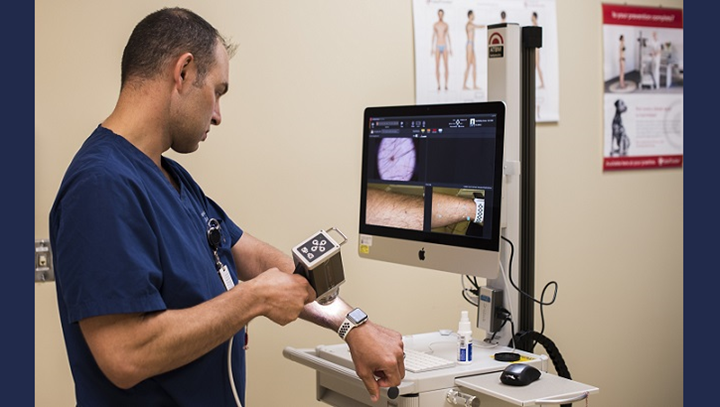
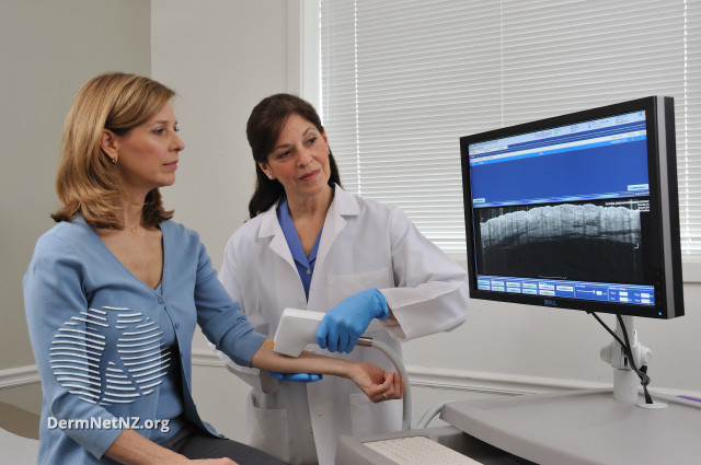
Over the past decade, Optical coherence tomography (OCT) has been increasingly used as a novel noninvasive imaging device to diagnose skin cancer. Its popularity is attributed to its ability to examine the skin in real time by providing a cross-sectional imaging of the skin. In comparison to older tools used by dermatologists such as the reflectance confocal microscopy, OCT has significantly better imaging depth of up to 2mm and a wider field of view albeit with a lower cellular resolution.

Various forms of OCT have since emerged such as Frequency domain OCT, dynamic OCT (D-OCT), and high-definition OCT (HD-OCT), all of which have been found to be efficacious in the diagnosis, treatment planning as well as monitoring of non-melanoma skin cancers.

With any diagnostic tool, continuous development and assessment is essential to examine the potential of the tool in clinical scenarios. To examine the diagnostic potential and accuracy of a tool, the most important factors are its sensitivity and specificity. However, the accuracy of the OCT is a subject of debate among dermatologists as many have expressed concern that screening with OCT alone as a diagnostic tool may lead to false negatives and delayed diagnosis of skin cancer. With this concern in mind, a research was conducted that examined the diagnostic accuracy of OCT to diagnose skin cancer.

The chief objective of this study was to estimate the diagnostic accuracy of OCT in basal cell carcinomas (BCC) and actinic keratosis (AK) as well as differentiating these lesions from normal skin.

Study Methodology

The study prepared a set of 142 OCT images that met the predetermined selection criteria for image quality as the diagnosis accuracy was heavily dependent on image quality. The OCT images for the study were all obtained by a widely used and commercially available OCT machine. The OCT images contained a mixture of cases with diagnosis of AK, BCC and normal skin.

These images were then presented simultaneously to two groups of blinded observers. One group comprised of 5 dermatologists who were experienced in OCT-image interpretation and diagnosis with a minimum of one-year experience in using OCT. The second group consisted of 5 dermatologists who were unfamiliar with OCT as a diagnostic tool. Each group of dermatologists were asked to answer a standardized questionnaire based on the OCT images and their presumed diagnosis.

Results of The Questionnaire

In the first group of dermatologists who were experienced with OCT, the diagnostic accuracy was found to be at a satisfactory level for Basal Cell Carcinoma with a sensitivity of 86% to 95% and a specificity of 81% to 98%. Unsurprisingly, the second group of dermatologists were not as adept at OCT as a diagnostic tool for BCC with the experienced dermatologists exhibiting an overall higher diagnostic accuracy in comparison to the OCT inexperienced dermatologists.

Proper OCT training is essential early on during residency years for dermatologists to be well versed with OCT, and only then can OCT be an effective screening asset for skin cancer.

References

Olsen J, Themstrup L, De Carvalho N, Mogensen M, Pellacani G, Jemec GB. Diagnostic accuracy of optical coherence tomography in actinic keratosis and basal cell carcinoma. Photodiagnosis Photodyn Ther. 2016 Dec;16:44-49. doi: 10.1016/j.pdpdt.2016.08.004. Epub 2016 Aug 9. PMID: 27519350. Feature image from DermNet, https://creativecommons.org/licenses/by-nc-nd/3.0/nz/legalcode,

Categories
Uncategorized

A 45-Year Historical Cohort Study of Cutaneous melanoma in 14·9 million people


This article is for informational purposes only and does not replace professional medical advice.

In the Nordic countries, the incidence of melanoma has been rising at a gradual yet steady rate in the past century. Multiple smaller scale studies have previously reported an increase in age-adjusted incidence of cutaneous melanoma (CM) in the Nordic countries over the last 60 years. However, there have been few prospective population-based studies that examined the occupational variation in CM risk over a period of time. In order to understand and determine the variations in the incidence of Cutaneous melanoma, a large-scale cohort study was done beginning from 1961 to 2005 by the Nordic Occupational Cancer Study team.

The chief objective of this cohort study was to determine the occupational variation in Cutaneous Melanoma and the associated risk with various occupations.

Study Methodology

Researchers undertook a massive 45-year follow-up of a historical prospective cohort study beginning from 1961 to 2005. The information for the study was obtained by linking records between the population census and cancer registry data for Nordic residents between the ages of 30 and 64 years living in Denmark, Finland, Iceland, Norway and Sweden. The study stratified the occupations into 53 categories each of which was further designated into indoor, outdoor and mixed work to estimate sun exposure. Furthermore, the data also analyzed the socioeconomic status of the entire population to determine its role on cutaneous melanoma.

With this data, standardized incidence ratios (SIRs) were calculated for each category of occupation and socioeconomic status. The SIR was calculated by dividing the observed number of Cutaneous Melanoma cases by the expected number of cases. Data for this calculation was obtained using stratum-specific person-years and national Cutaneous Melanoma incidence rates. The higher the Standardized incidence ratio, the higher should be the risk for skin cancer in a particular occupation.

Study Findings

The study evaluated a total of 385 million person-years, among which 83 898 incident cases of Cutaneous Melanoma was found. The study found that men with outdoor work had a low SIR of 0·79 and men with indoor work had a high SIR of 1·09. The differences in women based on their occupation also found similar results as in the men with higher SIR. In the group categorized under high socioeconomic status, the risk for melanoma was found to be even higher based on their SIRs which was found to be 1.34. in men and 1.31 in women. Likewise, people who worked in military, transport, technical, and public safety workers with substantial skin exposure to carcinogens had higher risk for skin cancer.

Conclusion

The major flaw in the study was the use of SIRs to estimate skin cancer risk as the denominator in the SIR equation was based on an expected number of cases which can be substantially higher when wrongly estimated skewing the equation to the wrong end. In people with outdoor occupation, there was reportedly a higher number of cases of melanoma in comparison to indoor worker but due to the observed cases being higher than the expected cases their SIRs were calculated as low. However, the study did find that occupational and socioeconomic variation in melanoma played a significant role in terms of risk for developing this fatal condition.

References:

Alfonso JH, Martinsen JI, Weiderpass E, Pukkala E, Kjærheim K, Tryggvadottir L, Lynge E. Occupation and cutaneous melanoma: a 45-year historical cohort study of 14·9 million people in five Nordic countries. Br J Dermatol. 2020 Jul 6. doi: 10.1111/bjd.19379. Epub ahead of print. PMID: 33026672.

Categories
Uncategorized

Is Self Teledermoscopy a Useful Diagnostic Tool For Skin Cancer?


This article is for informational purposes only and does not replace professional medical advice.

Skin cancer is one of the most prevalent forms of cancer in the world, with 16,221 cases being diagnosed annually in Australia alone. The highest risk factor for skin cancer is chronic exposure to UV rays that can cause mutations in DNA and mitochondrial genes. It is further divided into three types; Basal cell, Squamous cell, and melanoma. Of the three, melanoma is the deadliest, as it is responsible for a large proportion of skin cancer-related deaths despite accounting for only 2% of all cancers.

According to the Cancer Council, approximately 2 out of 3 Australians are diagnosed with skin cancer before they turn 70. Recent developments made in the field of skin cancer research have made early diagnosis much easier. Diagnosis of Skin Cancer for any skin pathology involves an initial suspicion for skin cancer due to positive family history and physical examination, after which, definitive diagnosis is made via skin biopsy and histopathological examination.

What is Teledermoscopy?

When a new patient visits a dermatologist for a skin lesion, the dermatologist’s initial step is to examine the lesion with his naked eye. To this end, a device called a dermoscope was designed that facilitates the diagnosis of skin lesions via usage of a magnifying glass and a light source to enhance the visualization of skin lesions in an outpatient setting.

More recently, there has been growing interest in the efficacy of mobile applications that combine the photographic and telecommunication features of a mobile with a magnifying glass to examine skin lesions.

Because it involves the use of telecommunication devices, the device could enable doctors’ to provide consultations to patients living in rural areas eliminating the need for patients living in remote areas to travel long distances for dermatological consultation. But as applications such as these tend to often result in incorrect self-diagnosis by patients, doctors are not quite convinced regarding the benefits of this tool.

Is Self Teledermoscopy equivalent to visiting a dermatologists?

Some researchers who were quite intrigued about the potential clinical applications of teledermatology have recently conducted a study that examines the accuracy of mobile digital teledermoscopy for skin self-examinations.

A randomized, controlled trial done in Brisbane, Australia studied 234 participants who were divided into two groups, the control group, and the interventional group. The participants of the interventional group were provided with iPhone compatible dermatoscopes for conducting self-examinations. Both groups were then asked to perform self-examinations, the controlled group with the naked eye and interventional group with their provided dermatoscopes. Researchers found no significant benefit of either methodology over the other. In fact, the differences in the number of diagnoses between the groups were almost negligible.

The findings of the study revealed no added benefit with the use of teledermoscopy devices for self-examination by patients. The applications were instead found to have both lower sensitivity and specificity in diagnosing skin cancer when compared to naked eye examinations by a dermatologist.

In an online survey performed amongst dermatologists in Australia, researchers found that most medical personnel were open to using dermatoscopy in their practices. They even found it helpful, especially for lesion monitoring. However, they were not very keen on the concept of patients diagnosing themselves with self-assessment tools such as teledermoscopy. The reason for their concern was mainly due to the high risk of a patient discarding malignant skin lesions as being simply benign after self-examination. This can lead to cancer progression and untimely diagnosis of skin cancer when it is at an advanced stage, during which treatment efficacy can be extremely poor.

Although self-examination of skin lesions is still highly recommended by dermatologists, they do not recommend the use of teledermoscopy by patients for diagnosing skin lesions. The scope of self-examination using either a patient’s naked eye or tools such as teledermoscopy should be limited only to the identification of any suspicious skin lesions. Once a suspicious lesion is identified, it should always be followed by a consultation with a dermatologist to rule out skin cancer.

References:

Warshaw E, Greer N, Hillman Y, et al. Teledermatology for Diagnosis and Management of Skin Conditions: A Systematic Review of the Evidence [Internet]. Washington (DC): Department of Veterans Affairs (US); 2010 Jan.

Linares, Miguel A et al. “Skin Cancer.” Primary care vol. 42,4 (2015): 645-59. doi:10.1016/j.pop.2015.07.006

Janda, Monika et al. “Evaluating healthcare practitioners’ views on
store-and-forward
teledermoscopy services for the diagnosis of skin cancer.” Digital health vol. 5 6 Feb. 2019, doi:10.1177/2055207619828225

Monika Janda, Caitlin Horsham, Dimitrios Vagenas, Lois J Loescher, Nicole Gillespie, Uyen Koh, Clara Curiel-Lewandrowski, Rainer Hofmann-Wellenhof, Allan Halpern, David C Whiteman, Jennifer A Whitty, B Mark Smithers, H Peter Soyer, Accuracy of mobile digital teledermoscopy for skin self examinations in adults at high risk of skin cancer: an open-label, randomised controlled trial, The Lancet Digital Health, Volume 2, Issue 3, 2020, Pages e129-e137, ISSN 2589-7500.

Categories
Uncategorized

Advancements in Treatment of Melanomas Over The Past 10 Years


This article is for informational purposes only and does not replace professional medical advice.

The prevalence and incidence of melanoma has increased exponentially in the past 20 years. Nevertheless, with significant advances in medicine over the past decade, almost 85% of melanoma patients have a good prognosis owing to early diagnosis and prompt management. 

Prior to advancements in the currently employed modes of therapy such as targeted therapy and immunotherapy, the primary agents used for treatment of melanoma was chemotherapy drugs such as dacarbazine. However, these therapies are far from ideal for patients due to their poor therapeutic benefits and associated adverse effects in advanced stages of melanoma.

Studies That Changed How Melanoma is Treated

Targeted Therapy

In order to find alternative treatment modalities, researchers focused on studies that strived to understand the underlying pathophysiology of melanoma. 

As researchers noticed the differences in the pattern of melanoma lesions in areas that were chronically exposed to sun against nonexposed areas, they shifted their attention towards genetic mutations resulting from damage by the sun’s harmful UV rays. 

Genomic sequencing of patients with chronic exposure to the sun versus those with limited sun exposure found unique mutation patterns in those with chronic sun exposure. This eventually resulted in the discovery of MAPK pathway which in turn led to the identification of BRAF and NRAS as the major gene mutations responsible for melanoma. 

With this newfound understanding of melanoma, targeted therapies were developed which was found to be significantly superior to chemotherapy in terms of both survival and adverse effects. 

Drugs such as vemurafenib, trametinib and dabrafenib that targeted BRAF mutations underwent clinical trials, which reported these agents to have profound albeit temporary responses in melanoma patients. Significant reduction in tumor burden was reported for short periods in advanced cases of melanoma. 

Further studies focused on combination therapy using BRAF and MEK inhibitory drugs. This combination treatment resulted in overwhelming inhibition of MAPK pathway. With these groundbreaking findings, the US FDA approved trametinib drug in 2013 and combination drug therapy with dabrafenib/trametinib in 2015 for treatment of melanoma. 

Additionally, combining immunotherapy with targeted therapy resulted in even better prognosis in melanoma cases. 

Immunotherapy

Immunotherapy for melanoma is done using monoclonal antibodies Ipilimumab that inhibit CTLA-4, and Pembrolizumab/Nivolumab that inhibit PD-1 receptors.

 In patients with advanced stage melanoma, the first study that examined the effects of immunotherapy was conducted using low-dose Ipilimumab which reported an overall 11% improvement in overall survival. Follow-up of this study combined a slightly higher dosage of ipilimumab with the chemotherapeutic drug dacarbazine but found no increase in benefits with the higher dosage or the combination therapy. Hereafter, low dose ipilimumab was approved for treatment of melanoma. Ipilimumab was associated with some symptoms of toxicity such as hepatitis, diarrhea, hypophysitis and rash in almost 30% of treated cases. 

Subsequent studies that compared ipilimumab with pembrolizumab found the latter to be associated with greater one-year survival benefits as well as lower rates of adverse effects. Henceforth, the US FDA approved pembrolizumab as a treatment modality for melanoma in 2014 as a second-line treatment drug but it was upgraded as a first-line treatment drug in 2015. 

Further studies that employed combined therapy with ipilimumab and nivolumab in melanoma patients proved to be highly beneficial but was also associated with increased rates of adverse toxic symptoms. Currently, FDA recommends using combination immunotherapy of ipilimumab and nivolumab as a first-line treatment for melanoma

Over the past decade, profound achievements have been accomplished in the treatment of melanoma, a condition that just 10 years back was akin to a death sentence due to its extremely poor prognosis. 

References

R.L. Siegel, K.D. Miller, A. Jemal Cancer statistics, 2016

CA Cancer J Clin, 66 (2016), pp. 7-30

C. Robert, L. Thomas, I. Bondarenko, et al.Ipilimumab plus dacarbazine for previously untreated metastatic melanoma

N Engl J Med, 364 (2011), pp. 2517-2526

C. Robert, J. Schachter, G.V. Long, et al.Pembrolizumab versus ipilimumab in advanced melanoma

N Engl J Med, 372 (2015), pp. 2521-2532

J.D. Walchok, V. Charion-Sileni, R. Gonzalez, et al.Overall survival with combined nivolumab and ipilimumab in advanced melanoma

N Engl J Med, 377 (2017), pp. 1345-1356

C. Robert, B. Karaszewska, J. Schachter, et al.Improved overall survival in melanoma with combined dabrafenib and trametinib

N Engl J Med, 372 (2015), pp. 30-39